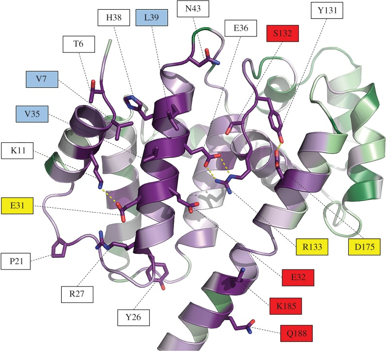
Figure 5.
Close-up view of the conserved residues of Psb29/THF1 identified by ConSurf analysis. The most conserved residues that are not buried within the Psb29 structure are shown in stick form, with red indicating oxygen atoms and blue nitrogen atoms. Intra-protein side-chain polar contacts are shown as yellow dashed lines. Some residues are colour-coded to indicate possible type of interaction. Red labels indicate potential hydrogen bonding/charged residues that might stabilize protein/protein interactions; yellow labels indicate residues possibly involved in both stabilizing the structure and interacting with proteins; light blue labels indicate potential hydrophobic contact sites.

