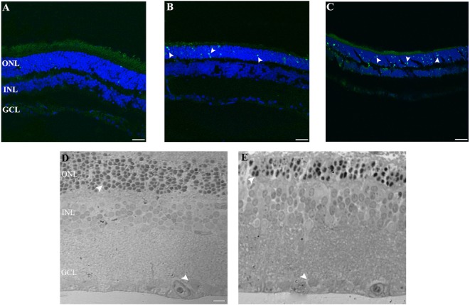
Figure 2.
Cell death study in retinal light damage: (A–C) TUNEL staining at 2, 4, and 8 days of LL treatment, arrowheads show the TUNEL positive in the ONL. (D,E) Transmittance images of entire width of retina maintained in LD (D) and LL8 treated (E), showing a clear reduction in ONL after 8 days of LL stimuli; normal (D) and pyknotic nuclei (E) in the ONL and normal nuclei in GCL (D,E). Scale bar indicates 30 µm. ONL, outer nuclear layer; INL, inner nuclear layer; GCL, ganglion cell layer.

