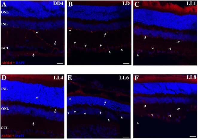
Figure 3.
OPN4 expression in retinal cuts of animals exposed to light: (A) retinas maintained in dark during 4 days showing positive labeling along the dendrites which extend into INL. (B) Retinas maintained in LD 8 showing a pattern similar to that in DD. (C–F) Light-treated animals during 1, 4, 6, and 8 days in LL showing increasing label restricted to cell soma around nuclear area. Arrow shows the positive labeling of OPN4 in soma, axons, and dendrites. The images are representative of three different experiments per treatment. Red: OPN4 antibody staining; blue: nuclear DAPI staining. Scale bar indicates 30 µm.

