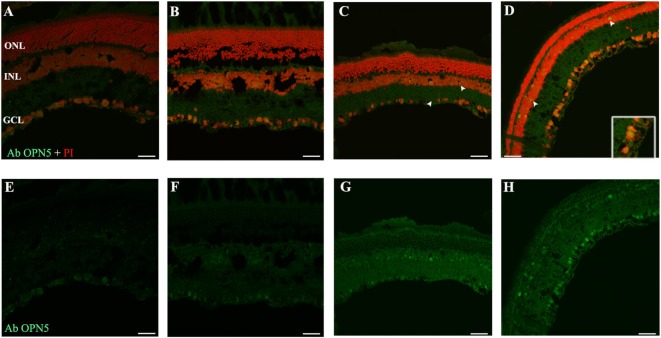
Figure 6.
Analysis of OPN5 protein expression in retina of animals exposed to light: (A,E) retinas maintained in dark during 4 days showing low positive staining in retinal ganglion cell (RGC). (B,F) Retinas maintained in LD 8 days showing a similar pattern as in DD. (C–H) Light-treated animals during 2 and 8 days in LL showing labeling in RGC and INL. The images are representative of three different experiments per treatment. Green: OPN4 antibody staining; blue: nuclear PI staining. Scale bar indicates 30 µm.

