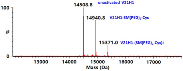
Figure 2.

Deconvoluted mass spectrum of the V21H1 antibody after activation by cross-linker and linkage to cysteine showing the distribution of non-activated antibody, antibody activated by one cross-linker, and antibody activated by two cross-linkers.
