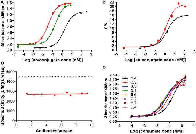
Figure 6.
(A) ELISA of biotin-V21H4 (black), V21H1-DOS47 (green), and V21H4-DOS47 (red) binding to recombinant VEGFR2. Results shown are representative of two to five experiments performed for each sample and are presented as the means and SE of samples tested in triplicate. (B) Binding of biotin-V21H4 (black) and V21H4-DOS47 (red) to VEGFR2 expressed by 293/KDR cells. Binding was quantified by flow cytometry. Results shown are representative of two to three experiments performed for each sample and are presented as the means and SE of samples tested in duplicate. (C) Urease enzyme activity of V21H4-DOS47 at different antibody/urease conjugation ratios (CRs). The dotted line represents unconjugated urease activity. (D) ELISA of V21H4-DOS47 with different antibody–urease CRs binding to recombinant VEGFR2/Fc. Results shown are representative of two experiments performed for each sample and are presented as the means and SE of samples tested in duplicate.

