
Fig. 2.
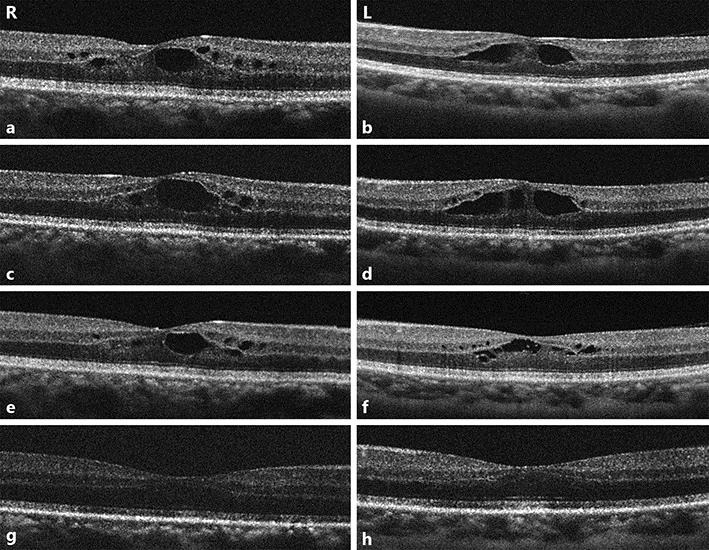
a–h Optical coherence tomography (OCT) scans of the macula densa of both eyes in time course. a, b OCT scans at diagnosis. Large cystic spaces are seen in the outer reticular layer and small cystic spaces in the inner nuclear layer. Retinal thickness is lower outside the perifoveal area. c, d OCT scans at 1 week following diagnosis, when paclitaxel was ceased. e, f One week after cessation of paclitaxel. g, h Three weeks after cessation of paclitaxel. Cystic spaces and retinal thickness gradually decreased.