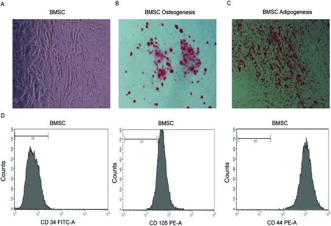
Figure 1. Morphology and identification of human BMSCs.
(A) Representative cell morphology of BMSCs at day 5. (B) Osteogenic differentiationof BMSCs, evident by Alizarin Red S staining. (C) Adipogenic differentiation of BMSCs, evident by Oil Red O staining. (D) Flow cytometry analysis of BMSCs. BMSCs were negative for CD34, and positive for CD105 and CD44. Magnification: 40× (A–C).

