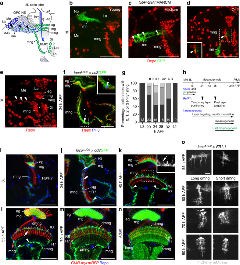
Fig. 2.
Development of astrocyte-like medulla neuropil glia. a Schematic of a third instar larval (3L) optic lobe. mng medulla neuropil glia, eg epithelial glia, GMC ganglion mother cell, La lamina, Lo lobula, Lop lobula plate, ln lamina neurons, Me medulla, meg medulla glia, mg marginal glia, mn medulla neurons, Nb neuroblast/neuroglioblast, OPC NE outer proliferation center neuroepithelium, R1-R6, R8/R7, R-cell axon subtypes, Xi inner chiasm glia. Arrowheads, migratory mng. b–d MARCM clones were generated using tubP-Gal4 and hs-FLP transgenes. Young GFP-labeled clones (green) closest to the OPC neuroepithelium contain Nb and neurons (b, n = 18). Medium (c, n = 6) and older (d, n = 11) clones include Repo-positive glial cells (arrowheads, red). e A projection of optical sections illustrates the migratory path of OPC-derived mng (arrowheads) through the cortex toward the anterior medulla neuropil edge (asterisk). f In 24 h APF optic lobes, mng were labeled with loco 1.3D2 -Gal4 UAS-cd8GFP (green) and Repo (red), and cells undergoing mitosis with phosphoHistone 3 (PH3, blue). g Percentage of optic lobes containing PH3-positive mng during early metamorphosis (n = optic lobes: 12, 23, 16, 18, 10, and 10). h Timeline of key steps underlying R-cell, target neuron, and glial development. i–n mng were labeled with loco 1.3D2 -Gal4UAS-cd8GFP (green) and Repo (blue). During 3L (i), at 24 h (j) and 42 h APF (k), mng cell bodies at the neuropil border (j, double arrowhead) migrate around the neuropil (j–l, arrowheads). Distal mng (dmng) extend short processes into the cortex at 42 h APF (k, insert) and into the neuropil from 55 h APF onwards (l, arrows). These become highly branched at 70 h APF and in adults (m, n). o Long and short dmng variants were labeled with UAS-FB1.1 (white). mng extend filopodia-like processes into the neuropil at 55 h APF. Primary processes project to final layers at 60 h APF. Increasingly branched secondary processes arise at 60, 70, and 80 h APF. Some dmng send thin processes along axon tracts into the cortex (arrow at 70 h APF). Panels f, i–n show single optical sections, b–e, o projections. For genotypes and sample numbers, see Supplementary Table 1. Scale bars, 50 μm (b–f, i–n), 10 μm (o)

