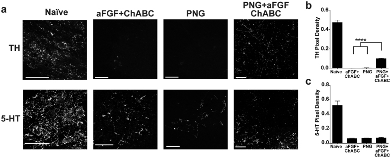
Figure 6.
PNG+aFGF+ChABC increases lumbar TH+ axons around the anterior horn. (a) Representative confocal images of TH+ (top row) or 5-HT+ (bottom row) stained transverse sections in the ventral grey horn of the lumbar spinal cord 40 weeks post-injury. Scale bar, 100 µm. (b,c) Quantification of the density of TH+ (b) and 5-HT+ (c) immunoreactivity in the ventral grey horn of the lumbar spinal cord. n = 6 per group except naive n = 4. ****p < 0.0001. One-way ANOVA, Fisher’s Least Significant Difference post-hoc test. Data represent mean ± S.E.M.

