Figure 2.
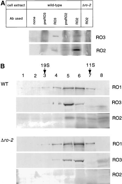
Association of N. crassa RO2 with RO3 (p150Glued). (A) RO2 and RO3 were immunoprecipitated in separate experiments with the use of anti-RO2 and anti-RO3 antibodies, respectively. Immunoprecipitants were resolved by SDS-PAGE, and RO2 and RO3 proteins were detected by Western blotting. The cell extracts and antibodies (antibody) used in these experiments are indicated above the immunoblot. (B) Cell extracts from wild type (WT) and Δro-2 were fractionated by centrifugation on 10-ml 5–20% linear sucrose gradients. Fractions (500 μl) were collected from the bottom of each tube (labeled lanes 1 through 8), and proteins were concentrated by trichloroacetic acid fractionation and then subjected to SDS-PAGE. RO1 (dynein heavy chain), RO2, and RO3 were detected by Western blotting. Sedimentation standards were thyroglobulin (19S), catalase (11S), and alcohol dehydrogenase (5S).

