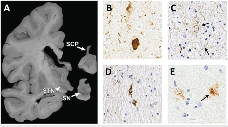
Figure 5-3.

Typical neuropathologic findings in progressive supranuclear palsy. The macroscopic photo shows atrophy of the superior cerebellar peduncle (SCP) and midbrain structures including the subthalamic nucleus (STN) and loss of pigmented cells in the substantia nigra (SN) (A).The photomicrographs show classic pathologic findings including neurofibrillary tangles (B), neuropil threads (C, arrows), coiled bodies (D), and tufted astrocyte (E, arrow) stained with PHF1 antibody to human tau.
Panel A is reprinted with permission from Dickson DW, et al, Curr Opin Neurol.18 © 2010 Lippincott Williams & Wilkins, Inc. journals.lww.com/co-neurology/Abstract/2010/08000/Neuropathology_of_variants_of_progressive.9.aspx.
