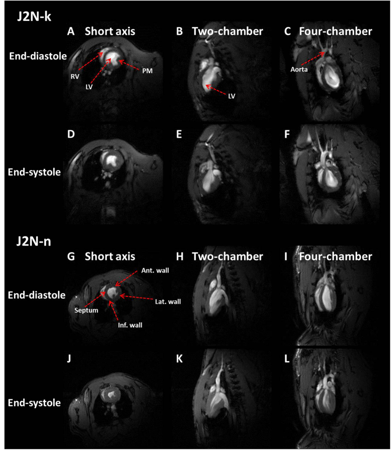
Figure 1.
Examples of typical three-plane images of J2N-k cardiomyopathic and J2N-n control hamsters obtained in three different views. (A,D,G and J) Short-axis view. (B,E,H and K) Two-chamber view. (C,F,I, and L) Four-chamber view. The red arrows indicate the left ventricle (LV), right ventricle (RV), aorta, papillary muscles (PMs), anterior (Ant) wall, inferior (Inf) wall, septum, and the lateral (Lat) wall.

