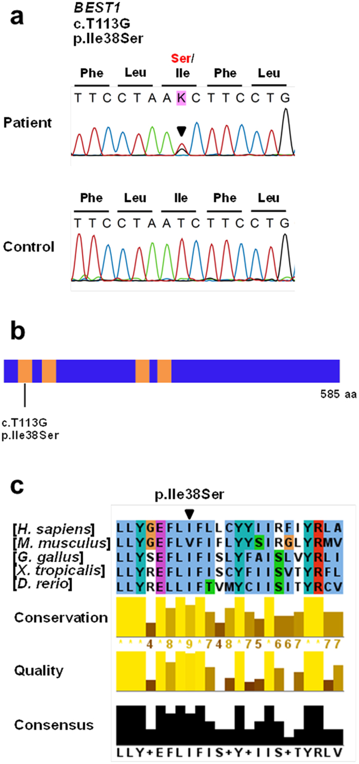
Figure 2.

Genetic analysis of the patient identified a novel BEST1 mutation, p.Ile38Ser. (a) Sequencing traces of the mutation and wild type control. Altered nucleotide and amino-acid changes are indicated above the sequence traces. (b) Domain structure of BEST1. The transmembrane (TM) domains are depicted by orange colored bars. The BEST1 mutation is located in the TM1 domain. (c) Partial protein alignment of BEST1 TM1 domain showed evolutionary conservation of the identified missense changes.
