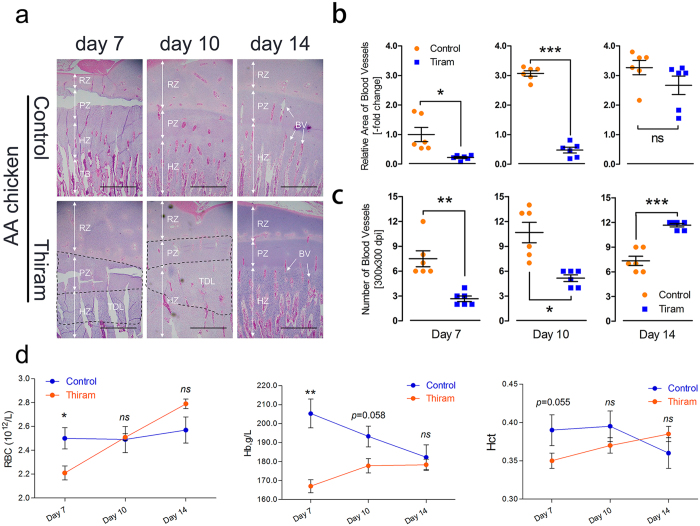
Figure 7.
Thiram suppresses tibial vascular distribution in the hypertrophic chondrocyte zones of AACs. (a) Tibial vascular distribution was significantly inhibited in the Thiram-treated group compared to the control group. The arrows indicate the BVs (blood vessels); TDL, tibial dyschondroplasia lesion; RZ, resting chondrocyte zone; PZ, proliferative chondrocyte zone; HZ, hypertrophic chondrocyte zone; and TB, trabecular bone. Scale bar = 500 µm. (b,c) The relative BV area and number were determined from two isolated groups using Image-Pro® Plus 6.0. Student’s t-test, * p < 0.05, ** p < 0.01, *** p < 0.001, n = 6; Error bars indicate SD. ns, not significant. (d) RBC (red blood cell) counts, Hb (hemoglobin) levels and Hct (hematocrit) values were determined for all blood samples. Student’s t-test, * p < 0.05, ** p < 0.01, n = 6; Error bars indicate SEM. ns, not significant.

