Figure 6.

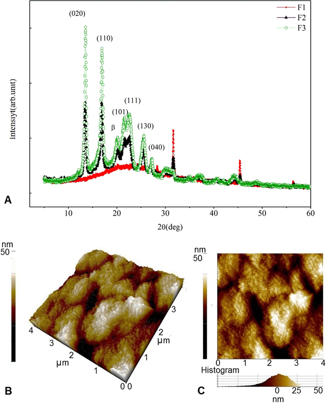
(A) X-ray diffractograms of the film. The peaks represented in green (F1) are for the film prepared using increased concentration of PHB, Nm and glycerol blend (2:1:1) with high intensity peaks, indicating the crystallinity of the film. Peaks shown in black (F2) correspond to the film prepared using equal proportion of the components Nm: PHB: glycerol (1:1:1) and displayed a similar diffraction pattern as the peak obtained with the increased concentration of PHB. Peaks represented in red (F3) are the film prepared using increased melanin: PHB: glycerol (2:1:1) which displayed a broader peak in the diffraction pattern of 20.2° to 26.8°. (B,C) Figure shows the AFM images of the samples scanned in an area of 4μm2. AFM topographical images of melanin nanocomposite film prepared with equal proportion of PHB, Nm and glycerol blend (1:1:1). The topographical scale of the images in 3D (B) and 2D (C) are shown in 50 nm scales. The roughness of the film was found to be 12.4 nm.
