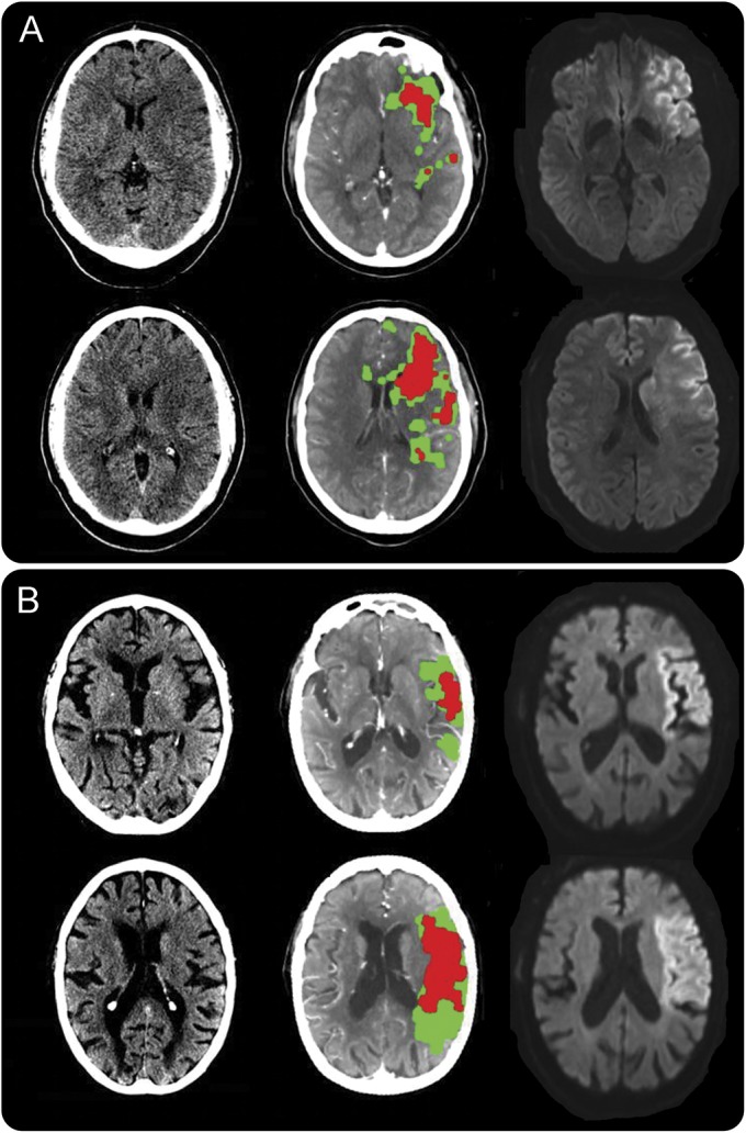
Figure 1. Noncontrast CT, core (red) and penumbra (green) CT perfusion maps, and acute diffusion-weighted imaging (DWI) MRI in 3 different patients.

(A) Agreement among all imaging modalities (Alberta Stroke Program Early Computed Tomography Score [ASPECTS] score 5, CT perfusion core volume 58 mL, MRI DWI volume 106 mL). (B) Established large infarct with high ASPECTS score (ASPECTS score 9, CT perfusion core volume 52 mL, MRI DWI volume 90 mL).
