
Figure 3.
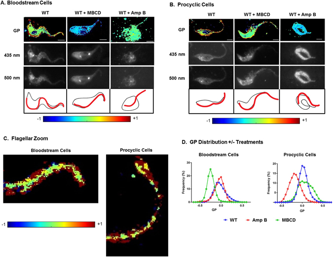
Sterol chelating agents disrupt membrane liquid order of bloodstream and procyclic cells differently. T. brucei bloodstream (A) and procyclic (B) cells were incubated with or without methyl-β-cyclodextrin (MBCD) or Amphotericin B (Amp B), stained with C-laurdan, and analyzed by fluorescence microscopy. Generalized polarization (GP) was calculated at each pixel in the two-dimensional image to determine the ratio of emission spectra obtained at 435 nm and 500 nm (see Methods). Higher liquid order (raft-enriched) is reflected by a higher GP (yellow-red), while lower liquid order is reflected by a lower GP (blue-green). The fluorescence images at the two wavelengths are shown in the middle panels and line drawings showing the cell body and flagellar membranes in black and red, respectively, are shown below these. Amp B treatment disrupts cell morphology but C-laurdan staining remains. MBCD has a milder effect on the cell morphology. The GP scale is provided at the bottom. Scale bar = 5 μm. (C) Higher power view of the anterior tips of untreated bloodstream and procyclic cells containing a thin cell body extension and mostly flagellum and having high GP. (D) Quantitative image analysis of individual pixels from cell images like those in (A and B) showing a reduction in liquid order in bloodstream cells by MBCD and in procyclic cells by Amp B from two biological replicates of procyclic cells and three biological replicates of bloodstream cells.