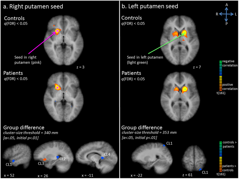
Figure 2.
Functional connectivity (Fc) maps for the putamen seed; panel a: right putamen seed, panel b: left putamen seed. Upper and middle row: Statistical maps resulting from the seed-based correlation analysis separate per group. Seeds are overlaid in pink (right putamen seed) or light green (left putamen seed). Maps are thresholded at q(FDR) < 0.05. Lower row: Group differences in functional connectivity per seed region (red indicates patients show increased connectivity as compared to controls, blue indicates patients show decreased connectivity as compared to controls).

