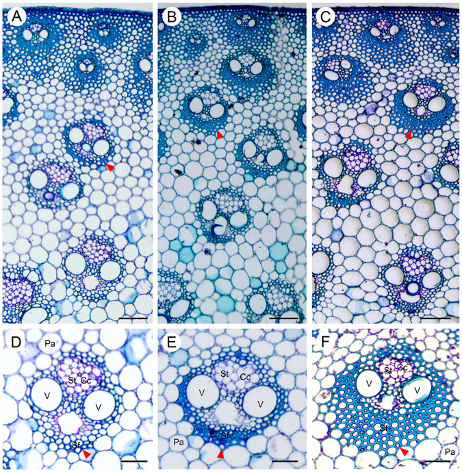
Figure 1.

Anatomy of an elongating internode of M. lutariorioparius. Sections from three segments of the 2nd internode from top were stained with Toluidine Blue O and observed under bright field. (A) upper internode (UI), (B) middle internode (MI), (C) basal internode (BI), (D,E and F) represent the magnified view of a vascular bundle in A, B, and C, respectively. Arrow head indicates the thickened fiber cells. Cc, companion cells, Pa, parenchyma, Sf, sclerenchyma fiber, St, sieve tube, V, vessel. Scale bar 100 μm in (A–C), and 50 μm in (D–F).
