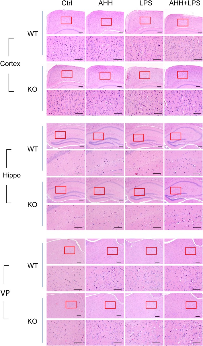
Fig. 3.
WIP1 loss worsens histological lesions in brain after hypoxic inflammation. Representative images of hematoxylin and eosin staining in the cortex, hippocampus (Hippo), and ventroposterior nucleus of the thalamus (VP) of WT and WIP1-KO mice (n = 10) after acute hypobaric hypoxia (AHH), LPS, and AHH + LPS exposure. Lower panels are enlargements of the enclosed area in the upper panels (scale bars, 200 μm).

