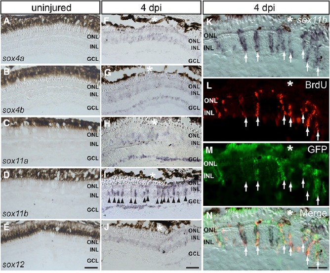
Fig. 3.
Spatial expression patterns of SoxC genes in the injured and intact retina. A–E In situ hybridization (ISH) of all SoxC genes on cryosections of the uninjured retina. F–J ISH showing the expression of SoxC genes in the injured retina at 4 dpi. K–N ISH of sox11b combined with BrdU/GFP immunofluorescence showing its expression in BrdU+/GFP+ cells in the INL at 4 dpi (arrows, co-localization of in situ, GFP and BrdU signals; *injury site; ONL outer nuclear layer, INL inner nuclear layer, GCL ganglion cell layer; scale bars 50 μm).

