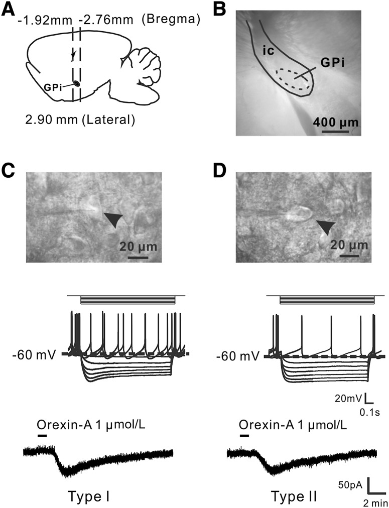
Fig. 1.
Orexin-A excites two types of GPi neurons. A Diagram of rat brain in sagittal view showing the location of the GPi between 1.92 and 2.76 mm from bregma. B A coronal brain slice containing the GPi. C, D Electrophysiological identification of Types I and II neurons in the GPi and the effect of orexin-A. The diameters of both types were >20 µm (arrowheads). Type I neurons showed a voltage sag during injection of a negative current step, whereas Type II neurons (n = 8, 10.5%) showed little or none. GPi, internal globus pallidus; ic, internal capsule.

