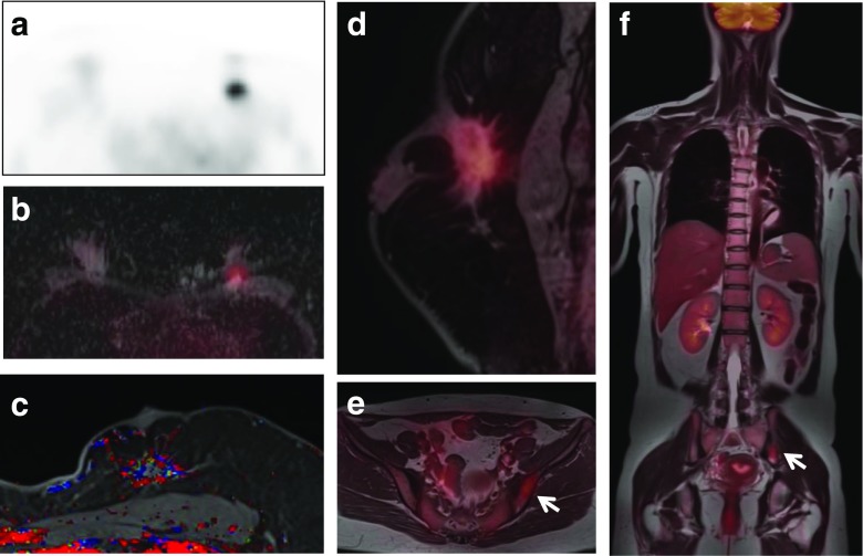
Fig. 1.
Images of whole-body FDG PET/MR scan and PET/MR mammography obtained with a dedicated four-channel PET/MR breast coil in a 41-year-old woman with invasive ductal carcinoma. A spiculated enhancing mass with strong FDG uptake, a low apparent diffusion coefficient (ADC) value, and a washout enhancement pattern is seen at the 12 o’clock position in the left breast on FDG PET (a), FDG/ADC PET/ mammography (b), color-coded map of the maximum slope of enhancement (c), and FDG/T1 fat-saturated gradient-echo (fs GRE) PET/MR mammography (d). Pelvic FDG/T1 and whole body FDG/T2 PET/MR images (e, f) show moderate FDG uptake in the left iliac bone suspicious for bone metastasis (arrows)

