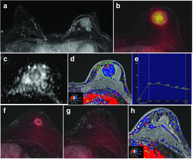
Fig. 3.
A 52-year-old woman with three invasive ductal carcinomas in the left breast. a The MIP of a contrast-enhanced T1 fs GRE subtraction image shows three enhancing masses in the left breast. b FDG/T1 fs GRE PET/MR mammography shows the largest hypermetabolic mass with a low ADC value (0.962 × 10⁻³ mm2/s) (c) and a washout enhancement (d, e). f, g FDG/T1 fs GRE PET/MR mammography shows two enhancing masses, one with moderate FDG uptake and one with no FDG uptake. h The smallest enhancing mass without FDG uptake is shown as a washout enhancement pattern on the color-coded map

