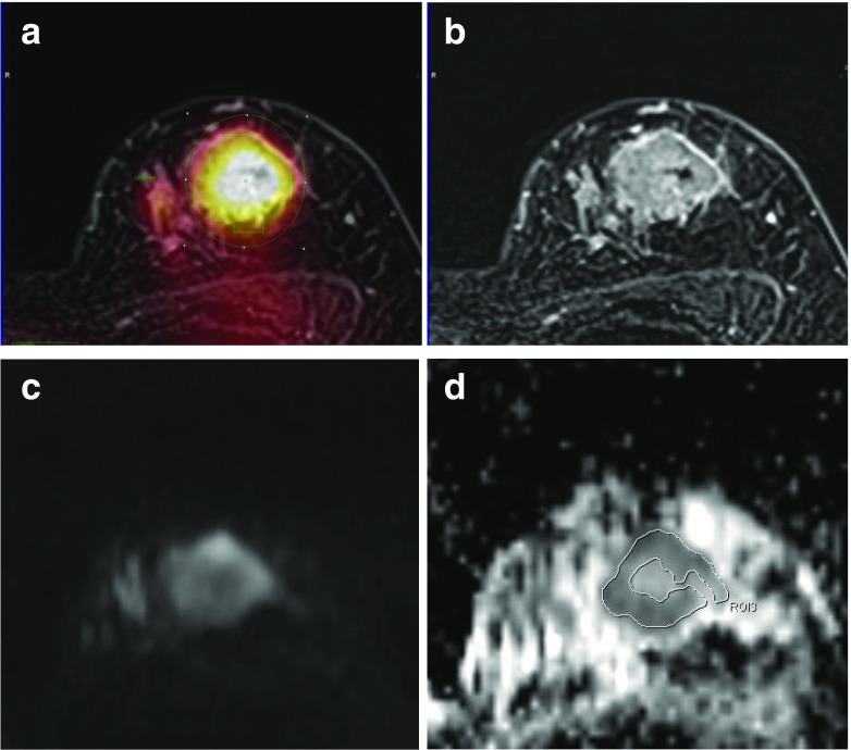
Fig. 8.
Comparison between FDG uptake and the ADC in a 52-year-old woman with a 3.5-cm invasive ductal carcinoma. a Region of interest for measuring SUVmax is drawn on the axial PET/MR mammography (SUVmax, 8.63). b Contrast-enhanced T1 fs GRE subtraction image showed a heterogeneous enhancing mass. c DWI (b value, 800 mm2/s) shows high signal intensity within the tumor. d For measurement of the mean ADC, a region of interest is manually drawn within a mass lesion on an ADC map (mean ADC, 0.825 × 10⁻³ mm2/s)

