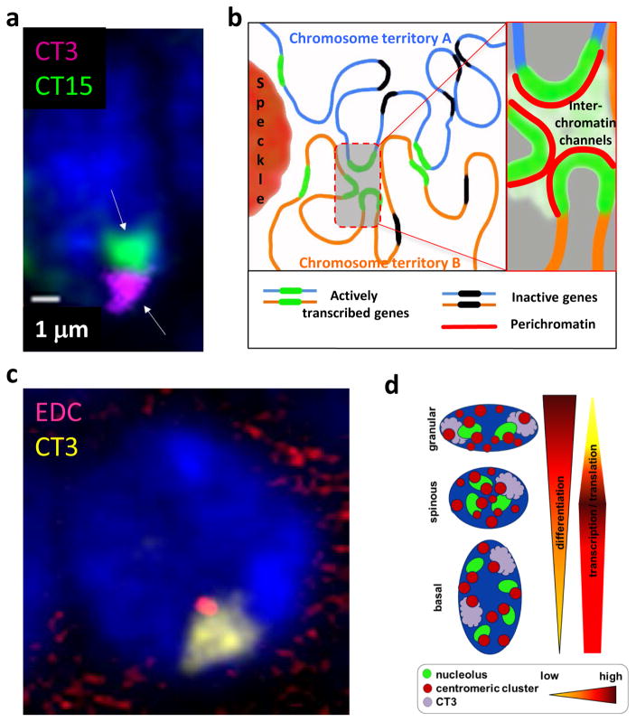
Figure 1. Changes in the spatial organization of the keratinocyte nucleus during epidermal development and differentiation.
A – 3D-FISH image of the nucleus of murine basal epidermal keratinocyte showing the positioning of the chromosomes 3 and 15 (arrows). Chromosome territory 3 (CT3, pink/violet) occupy more peripheral positioning in the nucleus, while the chromosome territory 15 (CT15, green) show more central positioning (courtesy of I. Malashchuk).
B - Chromosomes occupy distinct territories, in which distinct chromatin domains are permeated by interchromatin channels connected with a network of larger channels and lacunas separating distinct chromosomes and harboring different nuclear bodies including speckles (see Cremer et al., 2015, for details).
C – 3D-FISH image of the nucleus of murine basal epidermal keratinocyte showing the chromosome territory 3 (CT3, yellow) with EDC locus located at the internal part of the CT3 (red) (courtesy of I. Malashchuk).
D – Scheme illustrating the remodeling of 3D nuclear organization during terminal keratinocyte differentiation in the epidermis (see Gdula et al., 2013, for details).

