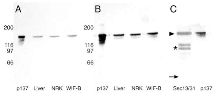
Fig. 2.
Anti-p137 antibodies detect a 170 kDa protein in rat liver, WIF-B cells, NRK cells and isolated yeast Sec13/13p complexes that comigrates with purified p137. Purified p137 (lane 1) and total homogenates of rat liver (lane 2, 20 μg), WIF-B cells (lane 3, 20 μg), and NRK cells (lane 4, 20 μg) were separated by SDS-PAGE (8–12% acrylamide) and subjected to immunoblot analysis using mouse anti-p137 (A) or rabbit anti-p137 (B) antibodies. Both antibodies detect a 170 kDa band that comigrates with purified p137. Purified yeast Sec13/31p complex and p137 were subjected to immunoblot analysis using rabbit anti-p137 (C). The antibody recognizes both p137 and yeast Sec31p (arrowhead), but not yeast Sec13p (arrow); the two additional bands detected are Sec31p degradation products (asterisk).

