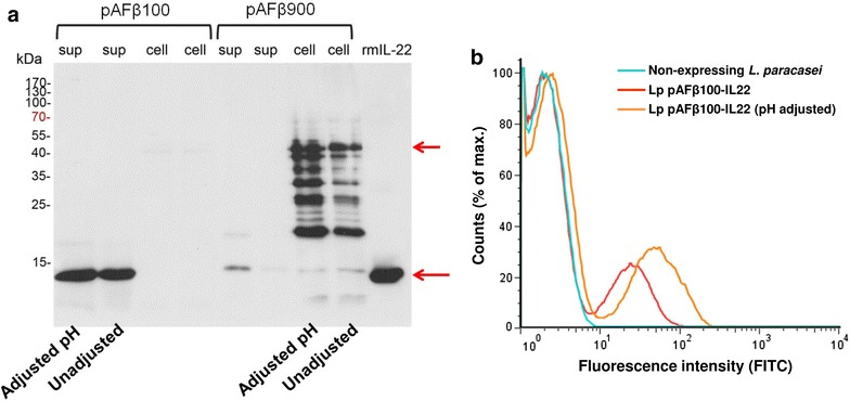
Fig. 2.

Expression and surface display of mouse IL-22 by L. paracasei BL23. a Detection of mouse IL-22 in the supernatant (sup) or cell pellet (cell) of Lp pAFβ100-IL22 (secreted-IL-22) and Lp pAFβ900-IL22 (anchored IL-22) by Western blot. b Surface display of anchored IL-22 (Lp pAFβ900-IL22) by flow cytometry. For both methods, the initial pH of culture MRS medium was either adjusted to pH 8.5 or not adjusted (pH 6.3)
