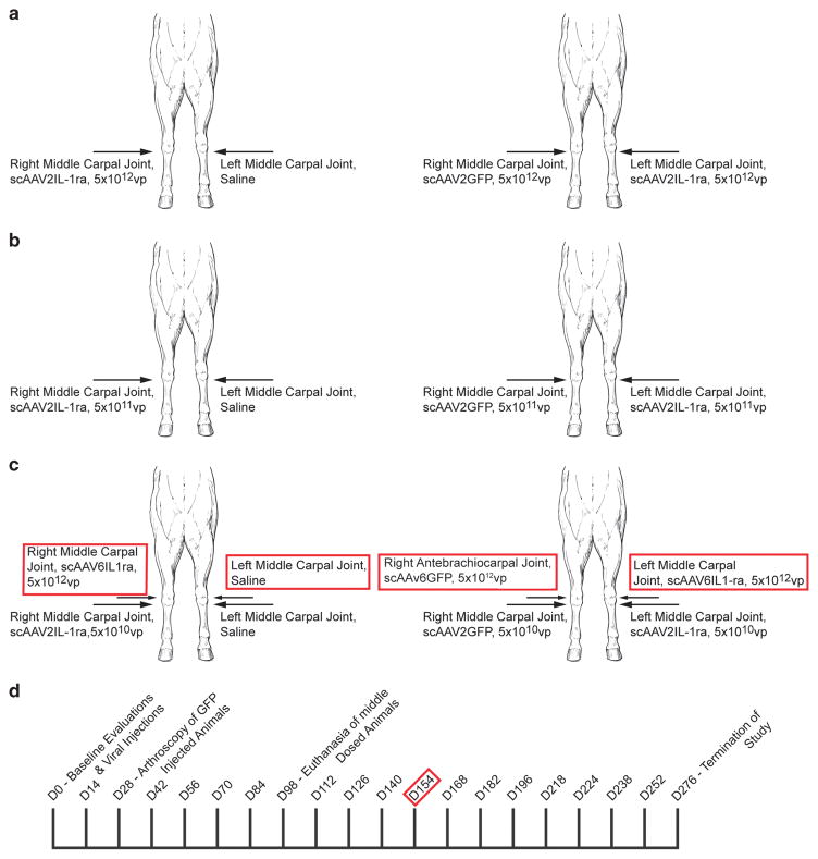
Figure 1.
Middle carpal joints were injected with saline, scAAV2GFP, scAAV6GFP, scAAV2IL-1ra and/or scAAV6IL-1ra as illustrated in panels a–c. Panel a represents the high dosed horses and the dosage of vector (or saline) and joint injected. Panel b represents the middle dosed horses and the dosage of vector (or saline) and joint injected. Panel c represents the low dosed horses and the dosage of vector (or saline) and joint injected. The red-bordered text represents what was injected on Day 154. The right antebrachiocarpal (verses the middle carpal) joint was injected with scAAV6GFP so that transduction could be determined from that particular vector and not be confused with previous GFP transduction from middle carpal joint injection. Panel d represents the time at which animals were dosed and/or redosed (red border), arthroscopically assessed and euthanized. Synovial fluid samples were collected for analysis at each time point listed.

