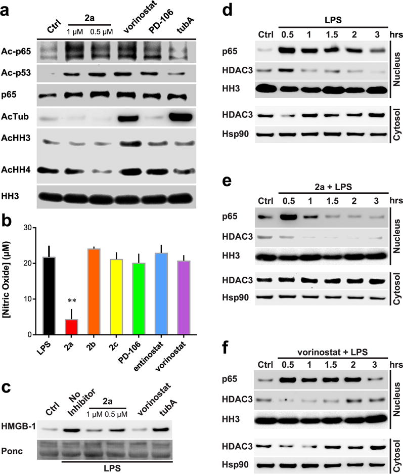
Figure 6.

Effects of HDAC inhibition on NF-κB p65 acetylation and inflammatory responses. (a) Western blot analysis of RAW264.7 cells treated with various HDAC inhibitors. Vorinostat and PD-106 used at 1 μM; tubA at 0.5 μM. (b) NO concentration secreted from RAW264.7 treated cells; normalized to control treated viable cell concentration. Molarity of 1 μM used for all inhibitors. n ≥ 3. (c) Western blot analysis of RAW264.7 cells. HMGB-1 secretion monitoring with ponceau stain as loading control. Cells were treated for 6 h. Molarity at 1 μM vorinostat and 0.5 μM tubA used. (d–f) Western blot analysis of LPS-treated RAW264.7 cells. Nuclear and cytosolic fractions split. 2a and vorinostat used to assess regulation of HDACs on Ac-p65 subcellular localization. Representative Westerns of n ≥ 2 experiments; error bars are SEM. **p-value < 0.0001.
