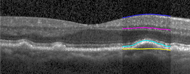
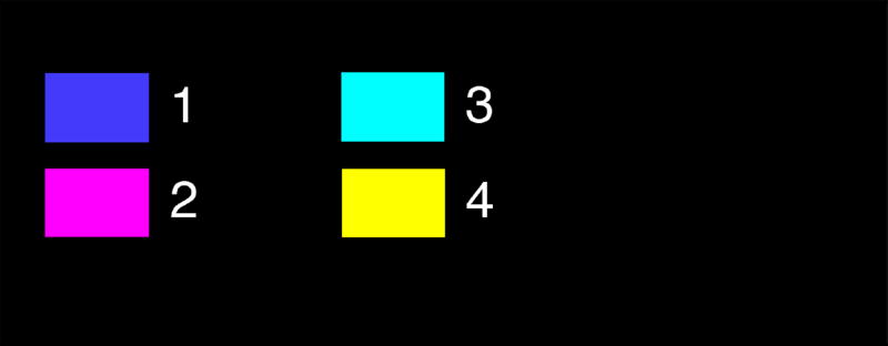
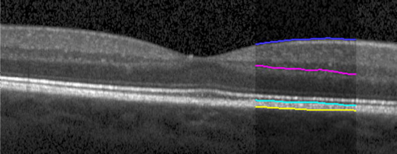
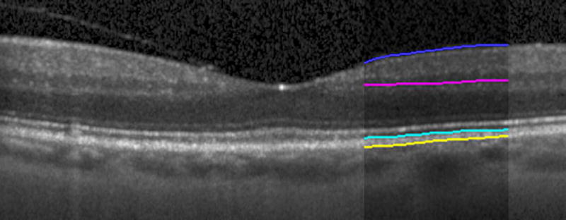
Figure 2.
Semi-automated segmentation of the inner retina, outer retina, and retinal pigment epithelium (RPE)-drusen-complex by Duke SDOCT Retinal Analysis Program V14.1.2. The inner retina is delineated from the internal limiting membrane to the inner aspect of the outer plexiform layer (between lines 1 and 2), while the outer retina is delineated from the inner aspect of the outer plexiform layer to the inner border of the RPE-drusen-complex (between lines 2 and 3). The RPE-drusen-complex extends from the inner aspect of the Retinal Pigment Epithelium plus drusen material to the outer aspect of Bruch’s Membrane (between lines 3 and 4), as seen in the color legend (Bottom right). Representative examples are shown for: No Apparent Aging (Top left), Early AMD (Top right), Intermediate AMD (Bottom left). Note the interdigitation zone is less apparent in the Early (Top right) and Intermediate AMD groups (Bottom left) than in the No Apparent Aging group (Top left).


