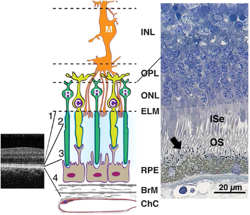
Figure 9.
Outer retinal anatomy relevant to interpreting retinal pigment epithelium (RPE)-drusen-complex thinning in early AMD. Abbreviations: C, cone; ChC, choriocapillaris; ELM, external limiting membrane; EZ, ellipsoid zone; INL, inner nuclear layer; ISe, inner segment ellipsoid; IZ, interdigitation zone; M, Müller cell; ONL, outer nuclear layer; OPL, outer plexiform layer; OS, outer segment; R, rod; RPE, retinal pigment epithelium.
SDOCT image of the perifoveal region in a normal macula (Left). The four outer retinal hyperreflective bands (from inner to outer between the expansion lines) are 1, external limiting membrane (ELM), 2, ellipsoid zone (EZ), 3, interdigitation zone (IZ), and 4, RPE-Bruch’s membrane complex. Contributory anatomical structures are expanded (not to scale) and schematized in middle panel. Cellular and extracellular components of the four outer retinal hyper-reflective bands include cones, rods, Müller cells, RPE, and Bruch’s membrane (Middle). Surrounding the photoreceptor inner and outer segments is a proteinaceous interphotoreceptor matrix (pale blue). Each individual photoreceptor is ensheathed by specialized domains (bright blue). Histology of outer retina from a normal human eye (Right, http://projectmacula). In this specimen, spindle-shaped melanosomes (arrow) can be recognized within RPE apical processes, interdigitated with the OS.

