
Figure 1. EWS-FLI1 changes localization upon trabectedin or lurbinectedin treatment.
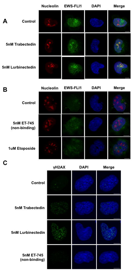
A) Single-cell imaging of HA-tagged TC32 cells treated with 5 nmol/L trabectedin or lurbinectedin for 6 hours shows EWS-FLI1 (green) localization into the nucleolus (red). DAPI (blue) was used as a nuclear stain. B) Single-cell imaging of HA-tagged TC32 cells treated with ET-745 or etoposide shows a lack of EWS-FLI1 nucleolar localization. C) Single-cell imaging of TC32 cells treated with 5 nmol/L Trabectedin, Lurbinectedin, or ET-745 for 6 hours shows the appearance of γH2AX (green) foci. DAPI (blue) used as a counterstain. Scale bars = 10 μm throughout this figure.