
Figure 4. Lurbinectedin synergized with SN38 to poison EWS/FLI1 activity and induced DNA damage.
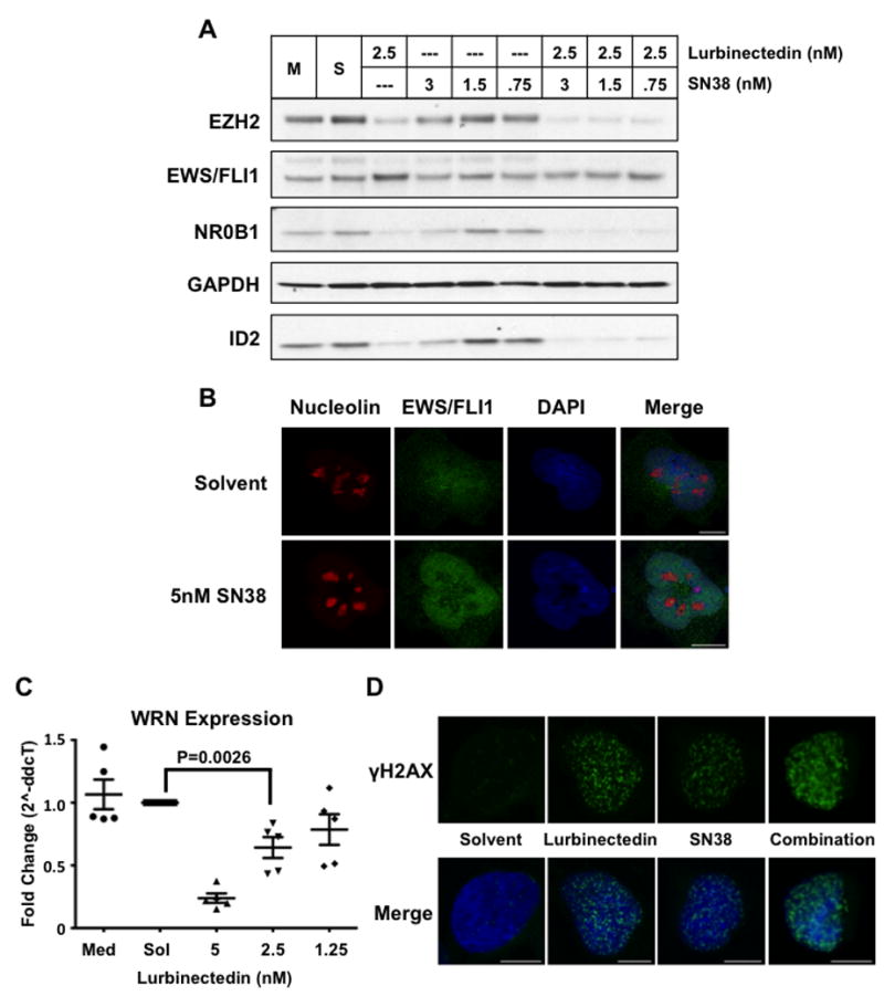
A) Western blot of TC32 cells after 18 h of treatment with either lurbinectedin alone, SN38 alone, or the combination at the indicated concentrations. B) Confocal microscopy of nucleolin (red) and EWS/FLI1 (green) in response to 5 nmol/L SN38 treatment after 6 h. DAPI (blue) staining of the nucleus. C) Quantitative PCR analysis of WRN mRNA expression in TC32 cells upon 12-h treatment with lurbinectedin at the indicated concentrations. D) Single-cell confocal microscopy showing γH2AX (green) foci upon 12-h treatment with 5 nmol/L of lurbinectedin, 5 nmol/L SN38, or the combination. DAPI (blue) staining of the nucleus. P-value was determined using a two-sided Student’s T-test. Scale bars = 10um throughout this figure.