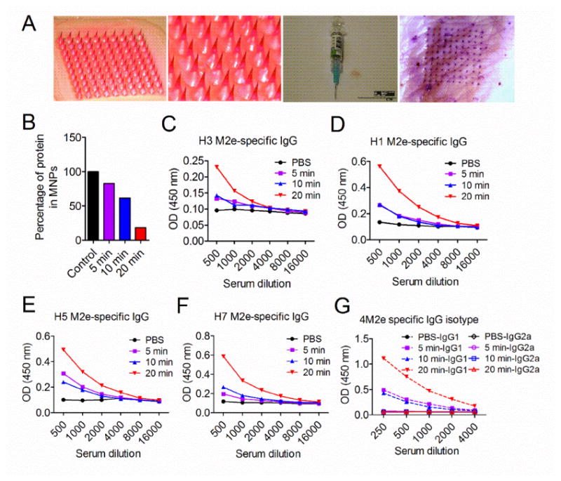
Fig 1.

4M2e-tFliC encapsulated MN patch induced antibody responses in mice. (A) 4M2e-tFliC was encapsulated into dissolving MNPs. An overview and zoom in view of sulforhodamine dye encapsulated MNPs (two panels in left). Contrast of the MNP and a traditional syringe needle (the third panel). View of porcine skin after administration with a purple dye encapsulated MNP (right panel). BALB/c mice were administered with 4M2e-tFliC MNPs using different application times: 5 min, 10 min and 20 min. (B) MNPs were retrieved after immunization and dissolved into ddH2O for remaining protein analysis. Sera were collected two weeks post immunization. The M2e specific IgG antibody responses (presented by OD values) were detected using different M2e peptides (the same as in 4M2e-tFliC) as ELISA coating antigens for antibody detection: (C) consensus H3; (D) H1; (E) H5; (F) H7. (G) M2e-specific IgG isotypes were detected using the pool of M2e peptides as coating antigens.
