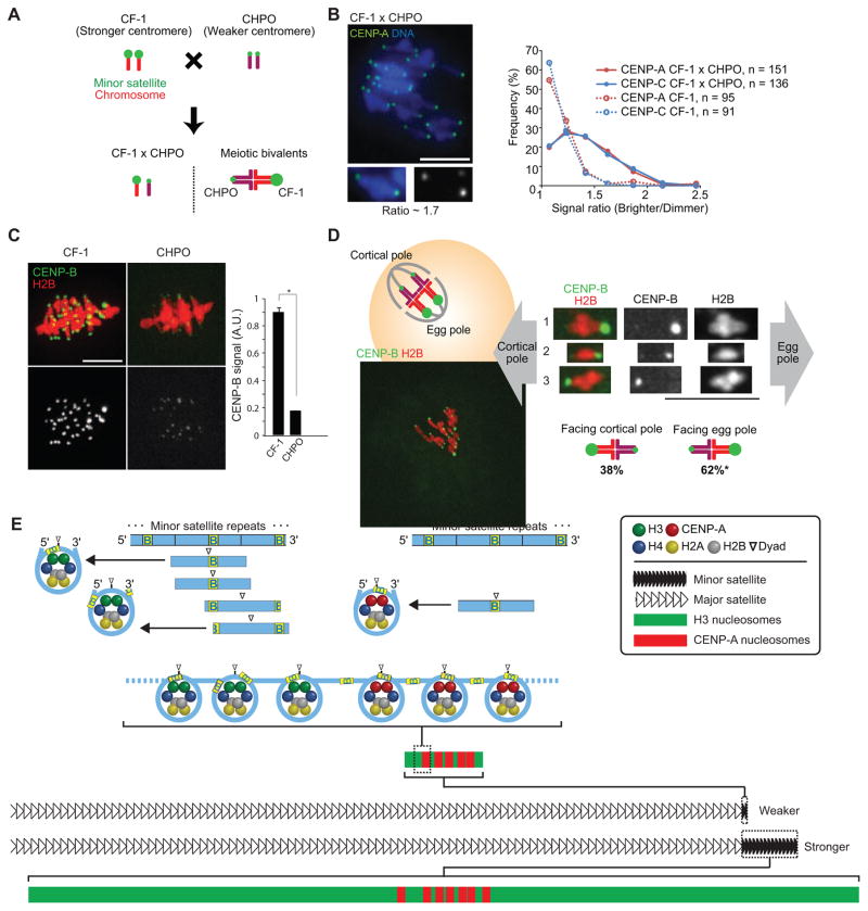
Figure 4. Stronger centromeres bind more CENP-A and CENP-C and orient preferentially to the egg in meiosis I.
(A) Progeny of a CF-1 × CHPO cross have meiotic bivalents with both weaker (CHPO) and stronger (CF-1) centromeres.
(B) CF-1 × CHPO oocytes were stained for CENP-A and CENP-C at metaphase I (see also Figure S4A–C). Image shows CENP-A staining, with a single bivalent magnified in the inset (arrows indicate paired centromeres). Graph is a histogram of CENP-A (red) and CENP-C (blue) intensity ratios, calculated as the brighter divided by the dimmer signal for each bivalent. CF-1 oocytes (dashed lines) are shown as controls.
(C) CF-1 or CHPO oocytes expressing CENP-B-mCherry and H2B-EGFP were imaged live at metaphase I. Graph shows quantification of CENP-B signals (mean±SEM, n≥340 centromeres from ≥27 oocytes in each case, pooled from three independent experiments). * p < 0.001; A.U., arbitrary units.
(D) Schematic shows bivalents in CF-1 × CHPO oocytes, with CF-1 centromeres facing the egg. Image shows a CF-1 × CHPO oocyte expressing CENP-B-EGFP and H2B-mCherry, shortly before anaphase onset; dashed white lines show cortex and spindle outline. The orientation of each bivalent was determined using CENP-B-EGFP intensity to distinguish CF-1 (brighter) and CHPO (dimmer) centromeres. * Significantly different from 50% (n=133, p<0.005). Images (B–D) are maximal intensity z-projections; insets are optical slices showing single bivalents. Scale bars, 10 μm.
(E) Model of our proposal that the amount of minor satellite determines centromere strength by constraining the spreading of CENP-A nucleosomes. Top: CENP-A nucleosomes (right) but not bulk nucleosomes (left) are strongly positioned on the minor satellite consensus, with the digestion-protected fragment centered on the last 10 bp of the 17-bp CENP-B box (yellow, labeled B). Major nucleosome positions are indicated by horizontal lines below the minor satellite consensus. Bottom: stronger centromeres contain more minor satellite DNA and centromeric proteins (e.g., CENP-A) than weaker centromeres. CENP-A nucleosomes localize to a small fraction of the minor satellite of stronger centromeres but occupy the length of the minor satellite DNA of weaker centromeres. CENP-A nucleosomes are shown with the dyad consistently positioned on the CENP-B box, whereas the position is variable for H3 nucleosomes (note that linkers between nucleosomes are drawn without CENP-B boxes for simplicity, and the linker length is drawn arbitrarily).

