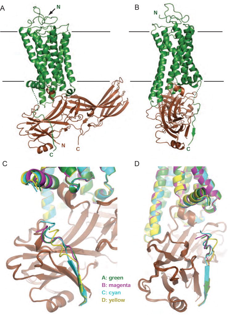
Figure 1.
Crystal structure of rhodopsin-arrestin complex with rhodopsin C-terminal tail interacting with arrestin N-terminal domain. See also Figure S1.
(A–B) Two views of T4L-rhodopsin-arrestin fusion complex with rhodopsin in green and arrestin in brown (T4L not shown). (C–D) Two views of rhodopsin C-terminal tails of overlayed four rhodopsin-arrestin complexes in an asymmetric unit. Complex A is shown in green, B in magenta, C in cyan, and D in yellow.

