
Fig. 2.
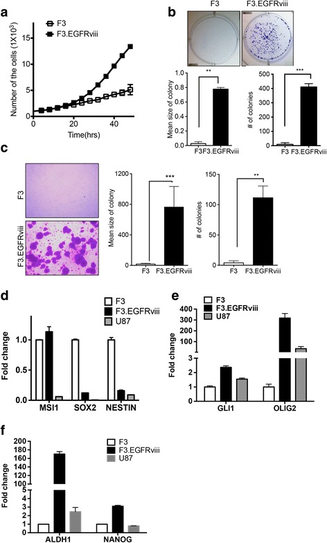
F3.EGFRviii cells as cancerous neural stem cells. a Accelerated proliferation of F3.EGFRviii cells relative to F3 cells was determined by cell counting at the indicated times. b Representative images from a clonogenic assay with F3 and F3.EGFRviii cells (top panel). The graphs show the mean number and size of colonies. c Images of colony formation in soft agar (left panel), and the colony number and mean colony size were presented (right panel). d-f The expression of neural stem cell and brain tumor markers in F3, F3.EGFRviii, and U87 cells was determined by real-time PCR (n = 3)