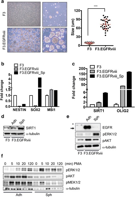
Fig. 3.

ERK1/2 activation in F3.EGFRviii spheres with increased cancer stemness. a Low- and high-magnification images of sphere formation in F3 and F3.EGFRviii cells after 14 days (left panels), and the mean sphere diameters from each cell type (n = 30, right panel). b and c Expression of the indicated genes in F3 cells, adherent F3.EGFRviii cells (Adh), and F3.EGFRviii spheres (Sph) was determined by real-time PCR (n = 3). d SIRT1 expression was determined by immunoblot analysis in F3 cells, adherent F3.EGFRviii cells (Adh), and F3.EGFRviii spheres (Sph) using α-tubulin as a loading control. e Immunoblot analyses of the indicated proteins in F3 cells, adherent F3.EGFRviii cells (Adh), and F3.EGFRviii spheres (Sph), with α-tubulin as a loading control (*, wild-type EGFR; arrow, expressed EGFRviii). f Adherent and spheroid F3.EGFRviii cells were treated with PMA (l μg/ml) and harvested at the indicated times for immunoblot analysis
