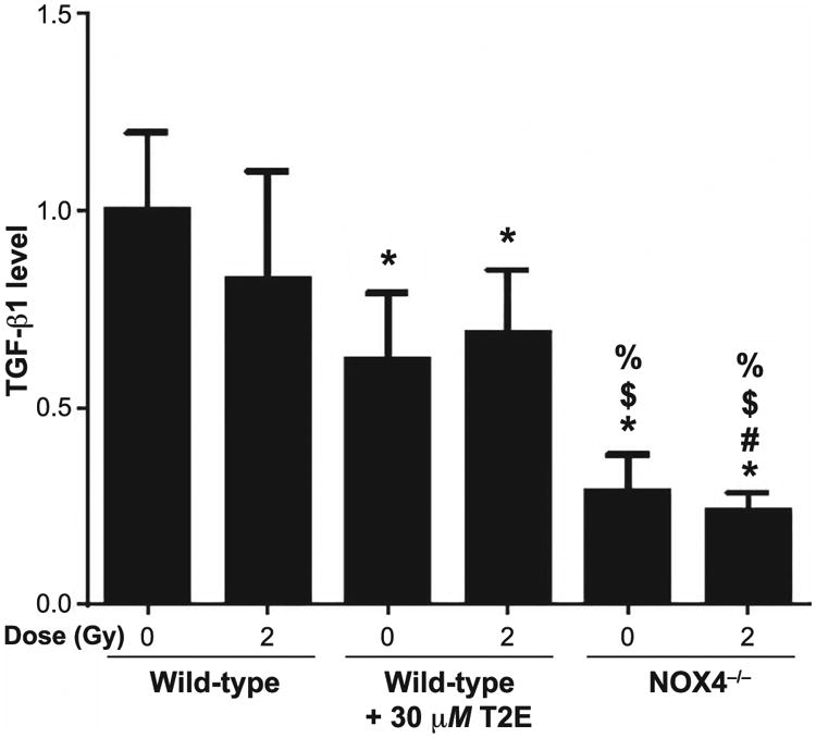
Fig. 7.

Measurement of extracellular TGF-β1. Conditioned media from PBS- or 30 μM MnTE-2-PyP (T2E)-treated wild-type mouse prostate fibroblasts and NOX4−/− mouse prostate fibroblasts were subjected to an activated TGF-β1 ELISA after activation of extracellular TGF-β1 by acidification. The data represent a fold change value of raw absorbance at 450 nm (subtracted by absorbance value at 540 nm). Data represent the mean ± standard deviation and were obtained from three independent experiments. The differences of mean absorbance were analyzed for significance using one-way ANOVA followed by post hoc Holm-Sidak correction for multiple comparisons. *Significant difference compared to wild-type nonirradiated fibroblasts; #significant difference compared to wild-type 2 Gy irradiated fibroblasts; $significant difference compared to T2E-treated nonirradiated fibroblasts; and %significant difference compared to T2E-treated 2 Gy irradiated fibroblasts.
