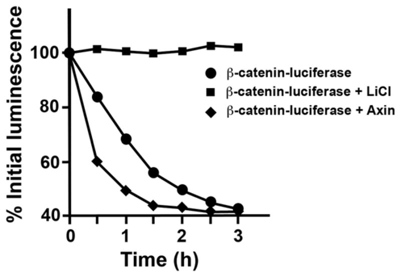
Fig. 3.

Reconstitution of β-catenin-luciferase degradation in Xenopus egg extracts. In vitro-translated β-catenin-luciferase fusion was incubated in Xenopus egg extracts in the presence of buffer, Axin (50 nM), or lithium chloride (20 mM; inhibitor of GSK3). At the indicated times, an aliquot was removed and luciferase activity measured. Background signal observed in the β-catenin-luciferase degradation assay is due to free luciferase protein, which degrades slowly
