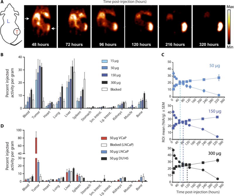
Fig. 1. 11B6 immunoPET imaging of PCa.

(A) Coronal slices through xenograft (LNCaP)-bearing mice over time. The long-lived PET isotope 89Zr enables longitudinal imaging, which shows continued uptake over 10 days. Schematic shows liver (L) and location of tumor (T) on flank. (B and C) Ex vivo determination (B) of organ and tumor antibody distribution at 320 hours, with time activity curves (C) in %IA/g of tumor (squares) and blood (circles) for 50-, 150-, and 300-μg doses (top to bottom). (D) Greater uptake in the higher hk2-producing VCaP in comparison to the LNCaP and nonproducing DU145 xenografts indicates specificity, which can also be blocked with cold antibody (1 mg). ROI, region of interest.
