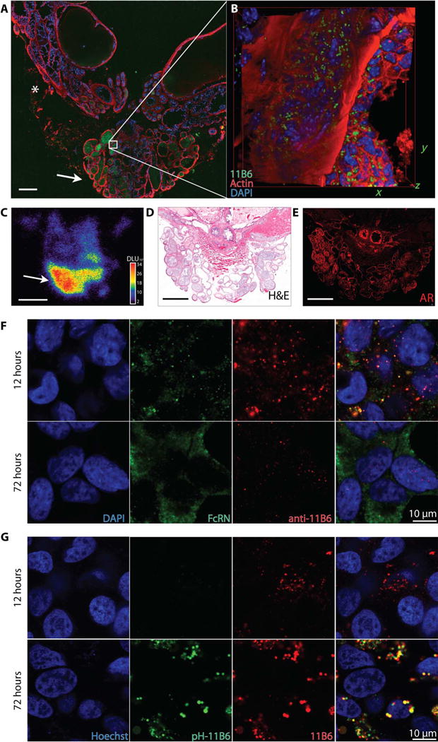
Fig. 3. Intracellular accumulation of 11B6-hK2.

The neonatal receptor facilitates internalization of the anti-hK2 immunocomplex. (A to C) The whole prostate and seminal vesicles (prostate package) were removed from Pb_KLK2 mice 72 hours after injection of Cy5.5-11B6 and 89Zr-11 B6 for whole-mount fluorescence (A), inset box volume scanned by confocal microscopy (B), and whole-mount autoradiography (C). Intense uptake was seen in the glandular structures of the ventral prostate (arrow), with lower uptake in the dorsolateral prostate (*). DAPI, 4′,6-diamidino-2-phenylindole; DLU, digital light units. (D) Radio and fluorescent signals in glands of the ventral prostate were localized by anatomical staining with hematoxylin and eosin (H&E). (E) AR staining is intense in the ventral prostate. Scale bar, 500 μm. (F and G) After incubation with LNCaP PCa cells, the 11B6 antibody colo-calizes with FcRn early (F) and is then trafficked to acidified lysosomes (G), as indicated by increased fluorescence from pH-responsive dye-labeled 11B6 (pH-11B6).
