Figure 1.
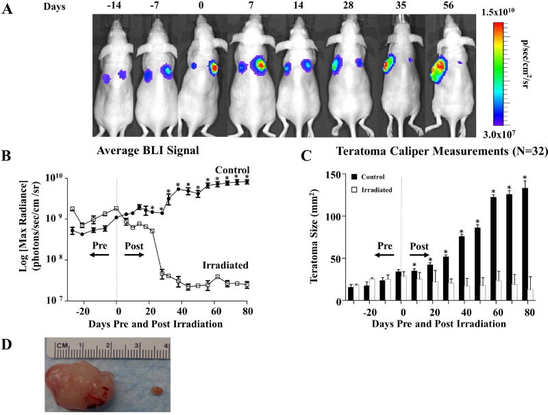
Irradiation arrests hESC-derived teratoma growth in vivo. (A) Representative BLI of teratoma growth and irradiation for immunodeficient mice seeded with 1×10⁶ H9 hESCs constitutively expressing FLuc-GFP on both dorsal flanks. At 28 days post-transplantation (day 0 pre-radiation), the larger of the two teratomas (right side) in this example was irradiated with 6 Gy daily for 3 continuous days (days 28–30 post injection) for a cumulative dosage of 18 Gy while the non-irradiated contralateral teratoma served as control as an un-irradiated control. A subsequent decrease in luciferase signal was observed on the irradiated side, whereas the non-irradiated teratomas demonstrated a progressively increasing luciferase signal. (B) Quantification of teratoma growth over time using BLI of luciferase signal from H9 hESC derivatives demonstrated growth arrest of irradiated tumors vs. control. (C) Changes in in vivo caliper measurements of teratomas over time. Non-irradiated teratomas increased in size over time, whereas irradiated teratomas decreased in size. (D) Explanted gross teratoma specimens from day 130 post seeding. Note the significant reduction in mass in the irradiated teratoma on the right compared to the non-irradiated teratoma on the left. *p <0.001.

