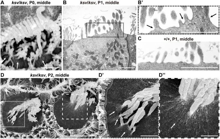
Fig 9. Deformation of the cuticular plate membranes of IHCs of ksv/ksv mice during the process of stereociliary fusion.
A. SEM image showing stereocilia in IHCs from the middle area of the cochlea in a ksv/ksv mouse at P0. Raised membrane of the cuticular plate (arrows). B. TEM image showing apical regions of the IHCs of a ksv/ksv mouse at P1. Highly magnified image of the stereociliary base shown in B’. C. Highly magnified TEM image showing the stereociliary base of the IHCs of a +/+ mouse at P1. D. Typical phenotypes of the apical surfaces of IHCs of ksv/ksv mice at P2. Highly magnified images in dotted and dashed boxes of D show the bulging of the cuticular plate membrane in D’ and D” (arrows), respectively. Scale bars = 3 μm (D), and 1 μm (A–C, D’ and D”).

