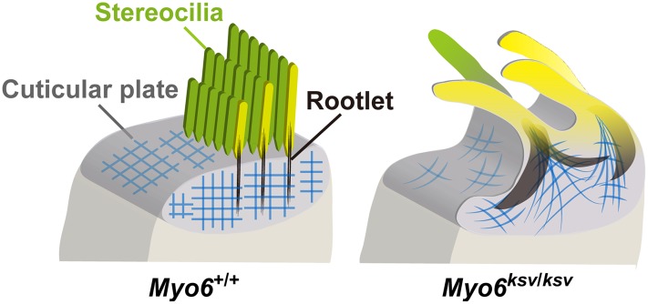
Fig 13. Schematic representation of the stereocilia in Myo6 mutant mice shows how stereocilia fuse.
Illustrations show apical surfaces of IHCs in +/+ (left) and Myo6 mutant (ksv/ksv) (right) mice as the model for the normal and abnormal architectures of the stereociliary base. In +/+ mice, stereocilia have dense rootlets (black) that extend through the taper region to anchor them into the actin mesh (blue) of the cuticular plate. The structures are maintained when MYO6 is normally expressed in the stereociliary taper, cuticular plate and cytoplasm (left), but the appreciable reduction of MYO6 by ksv mutations leads to stereociliary fusion accompanied by the deformation of the cuticular plates and the extension of the rootlets (right).

