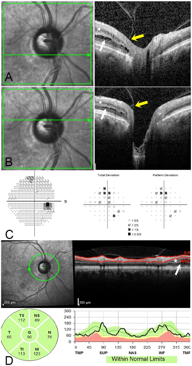
Fig 3. 54 year old male with pigmentary OAG with peripapillary retinoschisis and peripapillary atrophy.
(3A and 3B) Two horizontal OCT raster scans through the optic nerve head show splitting in the inner nuclear layer (white star) and outer plexiform layer (white arrows). There is a focal area of vitreopapillary traction at the temporal margin of the optic nerve head (yellow arrows). (3C) Humphrey visual field shows mild glaucomatous damage with a MD of -1.99 dB. (3D) Circumpapillary RNFL thickness map and section shows RNFL segmentation sparing the retinoschisis in the inner nuclear layer (white star), outer nuclear and outer plexiform layers (white arrow).

