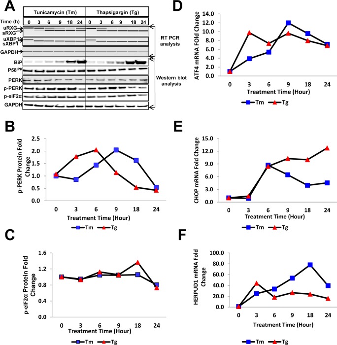
Fig 2. Validation of the RXG reporter system for the detection of cell stress by treatment with Tm and Tg by expression analysis of ER stress proteins.
(A) The kinetics of IRE1-mediated splicing of the synthetic reporter (sRXG) and endogenous XBP1 (sXBP1) were determined using RTPCR, followed by electrophoretic analysis (top panels) with GAPDH used as a loading control. Lysates from cells treated with Tm (10 μg/mL) and Tg (300 nM) were used for immunoblotting using antibodies directed against BiP, P58IPK, PERK, phosphorylated PERK, phosphorylated eIF2α and GAPDH. (B) Quantitation of p-PERK and (C) p-eIF2α proteins determined by Western blot analysis at 0–24 hours post cell treatments with Tm (10 μg/mL) and Tg (300 nM). (D) Kinetics of ATF4 and CHOP (E) mRNA activation measured by real-time qPCR following cell treatments with Tm and Tg. (F) Quantitation of HERPUD1 mRNA upon cell treatments with Tm and Tg.

