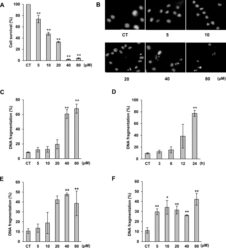
Fig 2. Effects of 3OTPCA on cell viability and DNA fragmentation.
(A) Percentage of viable cells in U937 cells treated with 0–80 μM 3OTPCA for 24 h. Cell viability was monitored using a CCK-8 assay. (B) Morphological change of nuclei was monitored by DAPI staining and fluorescence microscopy at 400× magnification. U937 cells were treated with various concentrations of 3OTPCA for 24 h. (C) DNA fragmentation in U937 cells treated with 3OTPCA for 24 h at the concentration indicated. (D) DNA fragmentation in U937 cells treated with 40 μM 3OTPCA for 3–24 h. (E-F). DNA fragmentation in Jurkat (E) and Molt-4 cells (F) treated with 3OTPCA for 24 h at the concentration indicated. The data represent the mean ± SD (N = 5). **p < 0.01 vs. CT (Student`s t-test).

